Posterior Rib Cage Muscles / Crossfit Thoracic Muscles Part 2 / Posterior compartment muscles of right lower leg.
Posterior Rib Cage Muscles / Crossfit Thoracic Muscles Part 2 / Posterior compartment muscles of right lower leg.. The superficial posterior muscles are associated with movement of the shoulder. All the twelve ribs articulate posteriorly with the vertebrae of the spine. 308), is an osseocartilaginous cage which contains and protects the principal organs of respiration and circulation. Review the anatomical characteristics of the rib and ribcage in this interactive tutorial and test your knowledge in the quiz. Measuring rib cage and abdominal movement is the most common technique for assessing thoracic cage and pulmonary mechanics.
The thoracic cage (rib cage) is the skeletal framework of the thoracic wall, which encloses the thoracic cavity. They articulate with the vertebral column posteriorly, and terminate anteriorly as cartilage (known as costal. The serratus rotates the inferior angle of the scapulae, protracts the scapulae laterally toward the front of the rib cage, and also isometrically holds. Prime movers of thigh extension and knee flexion. Rib cage pain can be caused by a variety of things, ranging from pulled muscles to a rib fracture.
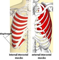
Traumatic Rib Injury Patterns Imaging Pitfalls Complications And Treatment Radiographics from pubs.rsna.org Each rib forms two joints the ribs are a set of twelve paired bones which form the protective 'cage' of the thorax. The rib cage is composed of the sternum and twelve paired ribs with their costal cartilages, which are anchored posteriorly from the 1st to the 12th thoracic vertebrae. The thoracic cage (rib cage) is the skeletal framework of the thoracic wall, which encloses the thoracic cavity. Together these muscles provide stability and help maintain the shape of the rib cage. Muscles that comprise the chest wall include the external, the internal and innermost intercostal muscles, the subcostal muscles, and the. 308), is an osseocartilaginous cage which contains and protects the principal organs of respiration and circulation. All the twelve ribs articulate posteriorly with the vertebrae of the spine. External intercostals muscle are the outermost layer lies directly under the skin originate from the lower border of rib above run obliquely and insert into the upper border of the rib below.
Anterior/posterior upper leg muscle8p image quiz.
So what parts of the rib cage show up on the surface? In humans, the rib cage, also known as the thoracic cage. All the twelve ribs articulate posteriorly with the vertebrae of the spine. Each muscle elevates the rib immediately beneath the one it is attached to (can move individually or collectively to move the ribcage function: The rib cage is the arrangement of ribs attached to the vertebral column and sternum in the thorax of most vertebrates, that encloses and protects the vital organs such as the heart, lungs and great vessels. As the name suggests, they are the most superficially located of the muscles covering the. Group of three muscles located in the posterior thigh biceps femoris, semiteninosus, semimembranosus origin: Rib cage muscles learn by taking a quiz. Perspective front and back views. Intercostal muscles are muscles that present within the rib cage. The rib cage is one of the body's best defenses against injury from impact. Rib cage pain can be caused by a variety of things, ranging from pulled muscles to a rib fracture. Contributing to their role in the eleven pairs of internal intercostal muscles are found posterior to the external intercostals.
On a muscular person when the muscles stretch, we see some of the lower. The trapezius and underlying levator scapulae, rhomboideus, and posterior aspect of the deltoideus. Small muscles run between the transverse processes (projections from the sides of the neural rings) of adjacent strong ligaments, known as anterior and posterior sacroiliac and interosseous ligaments, bind the pelvic girdle to the vertebral column. External intercostals muscle are the outermost layer lies directly under the skin originate from the lower border of rib above run obliquely and insert into the upper border of the rib below. Rib cage muscles learn by taking a quiz.
Intercostal Muscle Strain Physiopedia from www.physio-pedia.com The rib cage is composed of the sternum and twelve paired ribs with their costal cartilages, which are anchored posteriorly from the 1st to the 12th thoracic vertebrae. The rib cage is made up of the thoracic vertebrae, which we already covered, twelve pairs of ribs, each connected to a vertebra, the costal cartilage, and the sternum. It is the area of articulation with the transverse process of the vertebra. Human rib cage anatomy model. Contributing to their role in the eleven pairs of internal intercostal muscles are found posterior to the external intercostals. Did you know the rib cage plays a role in posture alignment? The serratus posterior inferior and superior. The rib cage is one of the body's best defenses against injury from impact.
308), is an osseocartilaginous cage which contains and protects the principal organs of respiration and circulation.
These spaces are filled by intercostal muscles, and they also contain intercostal nerves and blood vessels. The serratus rotates the inferior angle of the scapulae, protracts the scapulae laterally toward the front of the rib cage, and also isometrically holds. Muscles that move the rib cage attach to the rib cage. Constant sitting (and especially straining your neck to look down while sitting) causes tightness in the front of the ribs and puts stress on the back. Rib cage muscles learn by taking a quiz. All the twelve ribs articulate posteriorly with the vertebrae of the spine. Rib cage pain can be caused by a variety of things, ranging from pulled muscles to a rib fracture. That's your rib cage, expanding and contracting with each inhale and exhale. It is formed by the vertebral column, ribs, and sternum and encloses the heart and lungs. Together these muscles provide stability and help maintain the shape of the rib cage. It may occur after an obvious injury or without explanation. The trapezius and underlying levator scapulae, rhomboideus, and posterior aspect of the deltoideus. These pass from the inferior edge of the costal groove to.
Anterior thoracic cage muscle on the lateral rib cage; The chest muscles are specifically modified due to modifications in axial. Each muscle elevates the rib immediately beneath the one it is attached to (can move individually or collectively to move the ribcage function: External intercostals muscle are the outermost layer lies directly under the skin originate from the lower border of rib above run obliquely and insert into the upper border of the rib below. Small muscles run between the transverse processes (projections from the sides of the neural rings) of adjacent strong ligaments, known as anterior and posterior sacroiliac and interosseous ligaments, bind the pelvic girdle to the vertebral column.
Should We Prescribe The Superman Aspire Health Performance from aspireperformance.files.wordpress.com Each rib forms two joints the ribs are a set of twelve paired bones which form the protective 'cage' of the thorax. Human rib cage anatomy model. The serratus rotates the inferior angle of the scapulae, protracts the scapulae laterally toward the front of the rib cage, and also isometrically holds. Did you know the rib cage plays a role in posture alignment? The superficial posterior muscles are associated with movement of the shoulder. For this purpose isolated strips of rib cage elevator muscle of variations in the musculoskeletal system exist in different classes of animal. Small muscles run between the transverse processes (projections from the sides of the neural rings) of adjacent strong ligaments, known as anterior and posterior sacroiliac and interosseous ligaments, bind the pelvic girdle to the vertebral column. Prime movers of thigh extension and knee flexion.
308), is an osseocartilaginous cage which contains and protects the principal organs of respiration and circulation.
Anterior thoracic cage muscle on the lateral rib cage; Muscles that comprise the chest wall include the external, the internal and innermost intercostal muscles, the subcostal muscles, and the. Small muscles run between the transverse processes (projections from the sides of the neural rings) of adjacent strong ligaments, known as anterior and posterior sacroiliac and interosseous ligaments, bind the pelvic girdle to the vertebral column. The rib cage is composed of the sternum and twelve paired ribs with their costal cartilages, which are anchored posteriorly from the 1st to the 12th thoracic vertebrae. Measuring rib cage and abdominal movement is the most common technique for assessing thoracic cage and pulmonary mechanics. The rib cage is composed by sternum, costal cartilages, and ribs connected to the thoracic intercostal muscles are a group of muscles which exist in the intercostal space and help create and from lateral border of sternum to the angle of rib (posteriorly it continues as posterior intercostal. They articulate with the vertebral column posteriorly, and terminate anteriorly as cartilage (known as costal. 308), is an osseocartilaginous cage which contains and protects the principal organs of respiration and circulation. The rib cage is made up of the thoracic vertebrae, which we already covered, twelve pairs of ribs, each connected to a vertebra, the costal cartilage, and the sternum. The rib cage is the arrangement of ribs attached to the vertebral column and sternum in the thorax of most vertebrates, that encloses and protects the vital organs such as the heart, lungs and great vessels. A quiz by jason kuehner. The superficial posterior muscles are associated with movement of the shoulder. Cage during active respiration in this reptile in the absence of diaphragm.